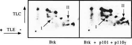
Figure 2.

Influence of PI 3-kinase-γ on the phosphopeptide map of Btk. Cells were radiolabeled with 32PO4, and Btk was immunoprecipitated and digested with trypsin as described in Materials and Methods. The phosphopeptides were separated by thin-layer electrophoresis and thin-layer chromatography and then visualized by autoradiography. Phosphopeptides I and II are indicated with arrows.
