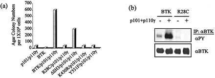
Figure 5.

Fibroblast transformation by PI 3-kinase-γ and Btk or Btk mutants. (a) Ten-thousand Rat-2 cells harboring SrcE378G and expressing PI 3-kinase-γ (p101 plus p110γ) and either wild type Btk or one of the Btk mutants (R28C, ΔSH3, K430R, or Tyr-551) were plated in duplicate into agar (6 cm). This figure does not show the colony number formed by each Btk mutant, which is the same as that formed by wild type Btk alone (less than 10 colonies). Colonies equal to or larger than 0.5 mm were counted and photographed 12 days postplating. Expression of p101, p110γ, and Btk was analyzed by immunoblots using anti-Btk or anti-myc epitope antibodies (data not shown). (b) Approximately 4 × 10⁶ Rat-2 cells expressing Btk, Btk/p101/p110γ, BtkR28C mutant, or BtkR28C/p101/p110γ were lysed, and Btk was immunoprecipitated using anti-Btk antibody. Immunoprecipitates were run on 8% SDS-PAGE gels and analyzed by immunoblotting with anti-phosphotyrosine (4G10) or anti-Btk antibodies, followed by either horseradish peroxidase-conjugated goat-anti-mouse or horseradish peroxidase-conjugated goat-anti-rabbit antibodies, respectively. Proteins were visualized by ECL.
