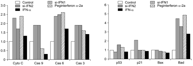
Fig. 5.
Change of pro-apoptotic molecules in tumor tissue treated with 50 × groups. Protein samples were extracted from the tumor tissues of control, si-IFN1, si-IFN2, peginterferon α-2a and IFN-α treated groups at 50 × concentrations. Protein sample were prepared for Western blot using antibodies to mouse cytochrome c, caspase 9, caspase 6, and caspase 3, p53, p21, Bax and Bad. Each band was further analyzed by densitometer. Each number on the figure represents the density compared control.

