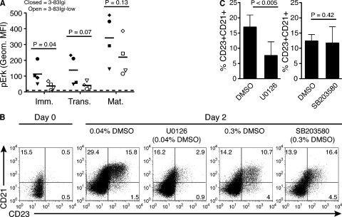
Figure 6.
Inhibition of Erk activity impairs the in vitro differentiation of normal immature B cells. (A) Bone marrow (immature) and spleen (transitional and mature) B cells from 3-83Igi and 3-83Igi-low mice were treated with pervanadate and analyzed by intracellular flow cytometry for expression of pErk. B cell subsets were defined as in Fig. 2 (C and D). Data represent the geometric MFIs from four mice and four individual experiments. Matching filled and open symbols represent mice that were analyzed at the same time (paired samples). The short black bars indicate average MFIs, and the dashed line just above the x-axis represents the average geometric MFI of isotype control staining. (B) Representative flow cytometric analysis of CD21 and CD23 expression in 3-83Igi immature B cells cultured as described in Fig. 5 A, with the exception that cells were treated with 10 µM U0126 (MEK inhibitor) in 0.04% DMSO, 10 µM SB203580 (p38 inhibitor) in 0.3% DMSO, or respective amounts of DMSO only during BAFF culture. Cells were analyzed at days 0 and 2 of BAFF culture. Numbers indicate frequencies of live B220+ B cells in quadrant gates. (C) Mean frequency (±SD; n = 3 mice from three individual experiments) of CD23+CD21+ 3-83Igi B cells at day 2 of BAFF culture as described in B.

