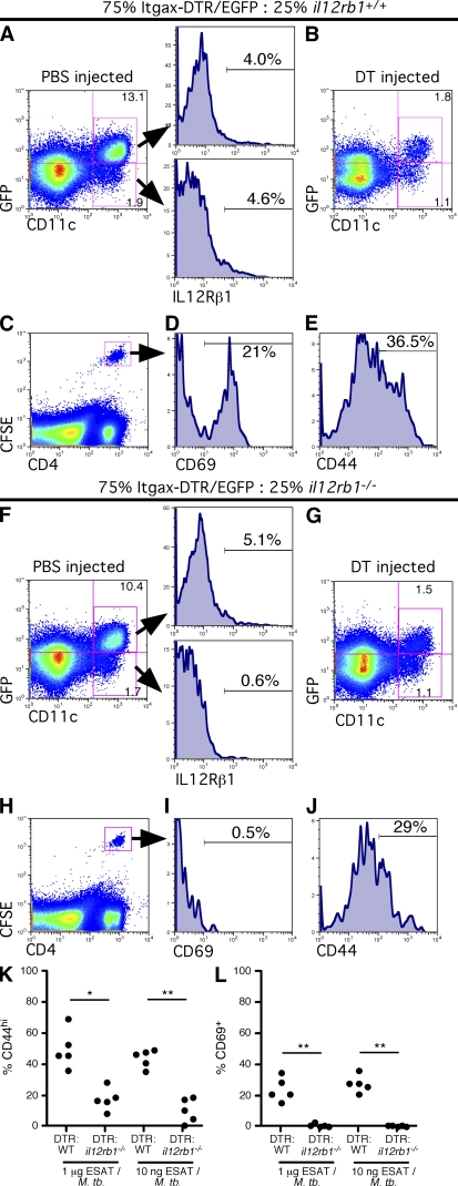
Figure 2.
The presence of il12rb1−/− DCs in the lung associates with impaired activation of M. tuberculosis-specific T cells in the draining MLN. Chimeras comprising 75% Itgax-DTR/EGFP:25% il12rb1+/+ or 75% Itgax-DTR/EGFP:25% il12rb1−/− were injected with either PBS (A and F) or DT (B–E and G–J). 12 h later, the frequency of CD11c+ GFP+ and CD11c+ GFP− cells remaining in the lungs after PBS injection (A and F) or DT injection (B and G) was determined. Gating based on CD11c+ GFP+ or CD11c+ GFP− cells demonstrated the level of IL-12Rβ1 surface expression (A and F). DT-injected mice subsequently received 1.5 × 10⁶ CFSE-labeled ESAT6-specific CD4+ T cells i.v. and 1 µg ESAT61-20/50 ng irradiated M. tuberculosis via the trachea. 12 h later, the frequency of CFSE+CD4+ cells in the draining MLN (C and H) and expression levels of the activation markers CD69 (D and I) and CD44 (E and J) determined by flow cytometry. The data points in K and L represent the CD44 (K) and CD69 (L) data from 5 mice per group that received either 1 µg or 10 ng ESAT1-20 peptide with irradiated M. tuberculosis and are representative of two separate experiments with 3–4 mice per group; for the difference in %CD44hi and %CD69+ ESAT-specific CD4+ cells between the indicated groups, *, P < 0.05; **, P < 0.005, as determined by Student’s t test.

