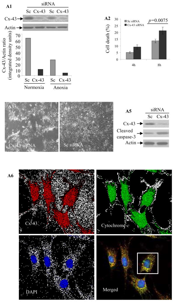
Figure 4.
(A1) Western blot of Sc siRNA (control) or Cx-43 siRNA transfected Sca-1+ cells grown under normoxia or OGD. Cx-43 siRNA successfully abolished Cx-43 expression. (A2) LDH release assay showing higher cell death in Cx-43 siRNA transfected cells as compared to Sc siRNA transfected cells under 4-h and 8-h OGD. Similarly, Cx-43 siRNA transfected cells (A3) showed more obvious rounded and shrunk morphology under 8-h OGD as compared to (A4) Sc siRNA transfected cells. (A5) Western blot showing elevated caspase-3 cleavage in Cx-43 siRNA transfected cells as compared to Sc siRNA cells. (A6) Double fluorescence immunostaining showing distribution of co-localized Cx-43 (red) and cytochrome-c (green) in mitochondria as punctate bodies in Sca-1+. (A7) The magnified image of a Sca-1+ cell from A6 (white box; original magnification=63x) showing clear apposition of red (Cx-43) and cytochrome-c (green). (B) Western blot showing higher level Cx-43 in the mitochondrial fraction of Sca-1+ after IGF-1 treatment as compared with cells without IGF-1 treatment. (C) IGF-1 treatment significantly increased Cx-43 level with concomitant suppression of Card-10 as compared to BSA treated cells. (D) Real-time PCR for mice sry-gene in female rat hearts (n= 4/group) on day-7 post-transplantation of male Sca-1+. The cells were transfected with Sc siRNA or Cx-43 siRNA. Cx-43 siRNA significantly reduced the survival of Sca-1+.


