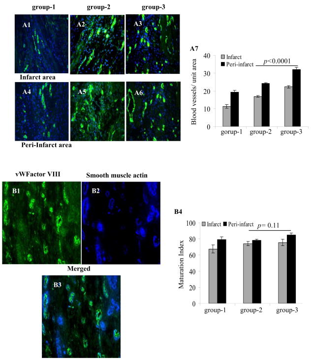
Figure 7.
Immunostaining of rat heart tissue sections for vWFactor-VIII (green) in the infarct (A1–A3) and peri-infarct (A4–A6) areas 6-weeks in group-1 (A1 & A4), group-2 (A2 & A5) or group-3 (A3 & A6). The nuclei were visualized with DAPI (blue). (A7) Blood vessel density was highest in group-3 in the infarct and peri-infarct areas. (B1–B4) Double immunostaining of rat heart tissue samples for vWFactor-VIII (green) and smooth muscle actin (SMA, blue) for determination of maturation index of the newly formed blood vessels in group-3. Blood vessel maturation index changed insignificantly between the three treatment groups. (Magnification=x400).

