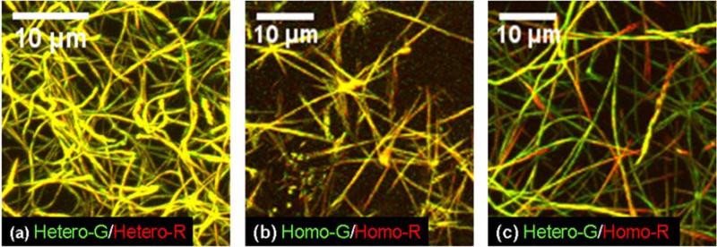
Figure 2.
3D reconstruction of confocal images of collagen fibril networks in different 0.3 mg/ml mixtures of fluorescently labeled wild type heterotrimers (Hetero) and oim homotrimers (Homo) from mouse tail tendons. Molecules labeled by AlexaFluor-488 (G) are shown in green color. Molecules labeled by AlexaFluor-568 (R) are shown in red color. The yellow overlay color appears in fibrils containing approximately the same amount of molecules labeled by each dye.

