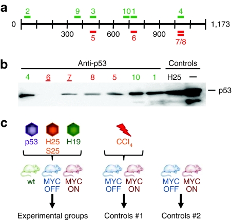
Figure 1.
Selection of anti-p53 shRNAs. (a) Illustration of the murine p53 mRNA (1,173 nt) and the location of the 10 different anti-p53 shRNAs used in this study. Colors indicate 19 mers (light) or 21 mers (dark). (b) Representative p53 western blot analysis from Huh-7 cells co-transfected with a p53 expression plasmid and a subset of the anti-p53 constructs (shRNAs 2, 3, and 9 were comparable to the other shown 19 mers). The two best shRNAs (6 and 7, both 21 mers) are underlined; they were later packaged into AAV-8 for analyses in mice. Control cells were co-transfected with the p53 plasmid and an unrelated shRNA (hAAT-25) or pBlueScript (last lane). (c) Injection schemes (see text for details). H19, hAAT-19 shRNA; H25, hAAT-25 shRNA; hAAT, human α-1-antitrypsin; nt, nucleotide; p53, anti-p53 shRNAs #6/7; S25, sAg-25 shRNA; sAg, surface antigen; shRNA, short hairpin RNA.

