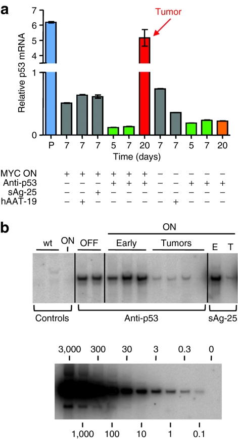
Figure 3.
Molecular analyses of AAV/shRNA-injected livers. (a) SYBR Green qRT-PCR analysis of p53 mRNA expression (mean ± SD), demonstrating reduction of p53 mRNA in both MYC ON and MYC OFF livers 5 and 7 days after p53 shRNA injection. Levels of p53 remained low in MYC OFF/p53 shRNA livers even 20 days after injection, whereas tumorigenesis in the MYC ON/p53 shRNA livers was associated with a significant p53 mRNA elevation. Lane P is a p53 plasmid-injected mouse liver used as positive control. (b) Southern blot analysis using a probe against a region conserved in all vectors. AAV DNA copy numbers (top panel) were high after initial injection of anti-p53 shRNA in MYC OFF livers (lanes “OFF”, each lane is an individual mouse) and MYC ON livers (lanes “ON Early”), as well as in MYC ON/sAg-25 livers (lane “ON E”). MYC ON/anti-p53 shRNA liver tumors (lanes “ON Tumors”) contained very little if any AAV DNA copies, identical to MYC ON/sAg-25 tumors (lane “ON T”). Lanes “Controls wt” are age-matched wild-type murine livers, and lane “Controls ON” is a noninjected MYC ON liver. The bottom panel shows a standard curve obtained using serial dilutions of the parental AAV/shRNA vector plasmid. AAV, adeno-associated virus; hAAT, human α-1-antitrypsin; qRT, quantitative real-time; sAg, surface antigen; shRNA, short hairpin RNA.

