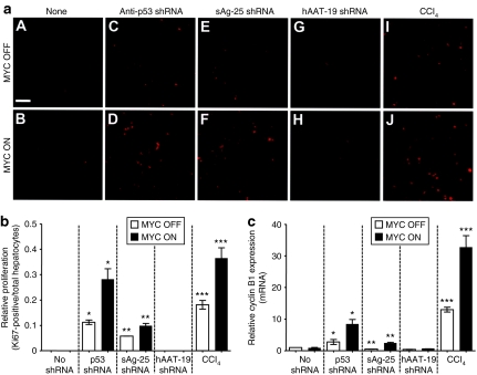
Figure 6.
MYC and shRNAs cooperate to induce hepatocyte proliferation associated with increased cyclin B1 expression. (a) Representative Ki67 immunofluorescence analyses of hepatocytes from livers treated with the indicated vectors in the absence (A, C, E, G, I) or presence (B, D, F, H, J) of MYC. Bar = 80 µm. (b) Quantitation of Ki67-positive hepatocytes in livers (treated as indicated) relative to total hepatocytes. (c) qRT-PCR analysis of cyclin B1 mRNA expression in the same livers. Cyclin B1 mRNA expression levels were normalized to ubiquitin expression levels and depicted relative (mean ± SD) to levels in a normal liver. Statistical significance was measured using a Mann–Whitney test (*P = 0.03, **P = 0.03, ***P = 0.03). AAV, adeno-associated virus; CCl4, carbon tetrachloride; HCC, hepatocellular carcinoma; qRT, quantitative real-time; sAg, surface antigen; shRNA, short hairpin RNA.

