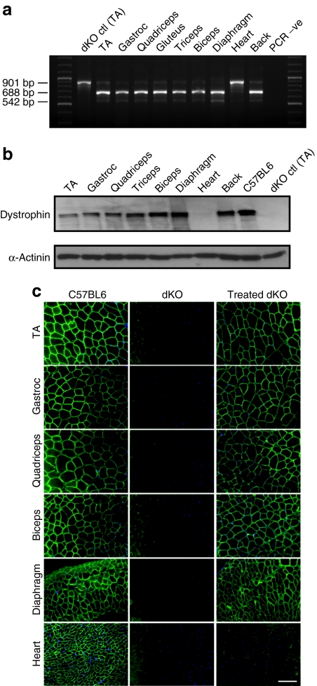
Figure 1.
Dystrophin expression in tissues from dKO mice following six once-weekly intraperitoneal injections of PPMO at 25 mg/kg/week. (a) Reverse transcriptase-PCR analysis to detect exon 23 skipping efficiency at the RNA level. The 901-bp product represents the full-length transcript, and the products of 688 and 542 bp represent transcripts that exclude exon 23 and exons 22 and 23, respectively. (b) Western blot to detect dystrophin expression in tissues from PPMO-treated dKO mice, compared with untreated dKO and C57BL6 control mice (top gel). Equal loading of 50-µg protein is shown for each sample with α-actinin expression detected as loading control (bottom gel). (c) Immunostaining of muscle tissue cross-sections to detect dystrophin expression and localization in C57BL6 normal control mice (left panel), untreated dKO mice (middle panel), and PPMO-treated mice (right panel; N = 5). Muscle tissues analyzed were from tibialis anterior (TA), gastrocnemius, quadriceps, biceps, diaphragm, and heart muscles. Bar = 100 µm. dKO, double-knockout; PPMO, peptide-conjugated phosphorodiamidate morpholino oligomer.

