
Figure 1.
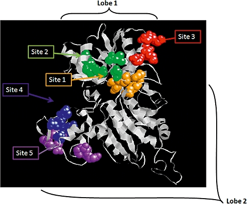
Molecular model of a CaR protomeric VFT domain. A model of a single subunit based on the mGlu-1 crystal structure 1EWK (Kunishima et al., 2000). Putative Ca2+o binding sites (1–5) were identified by aromatized terbium luminescence analysis of globular sub-domains (Huang et al., 2009). Site ‘1’ also corresponds to the conserved L-amino acid-binding site of class C GPCRs raising the possibility that Ca2+ and amino acid binding are closely associated. CaR, calcium-sensing receptor; mGlu, metabotropic glutamate receptor; GPCR, G-protein-coupled receptor; VFT domain, Venus Fly Trap domain.