Figure 3.

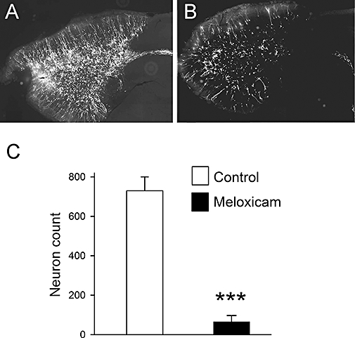
Effect of meloxicam on the appearance of new neurons in the OB. Six-week-old mice were treated with meloxicam (20 mg·kg−1) for 10 days. Then, 24 h after cessation of drug treatment, viral vectors encoding GFP were injected directly into the exit point of the SVZ adjacent to the origin of the RMS to label migratory neuroblasts. Two weeks later, animals were killed, and 40 µm sagittal sections obtained from the OB. The micrographs show GFP labelling at the level of entry of the RMS into the OB for a control animal (A) and a treated one (B). (C) Neuron counts in the OB, with each value being the mean determined from at least four animals. Bars show SEM.
