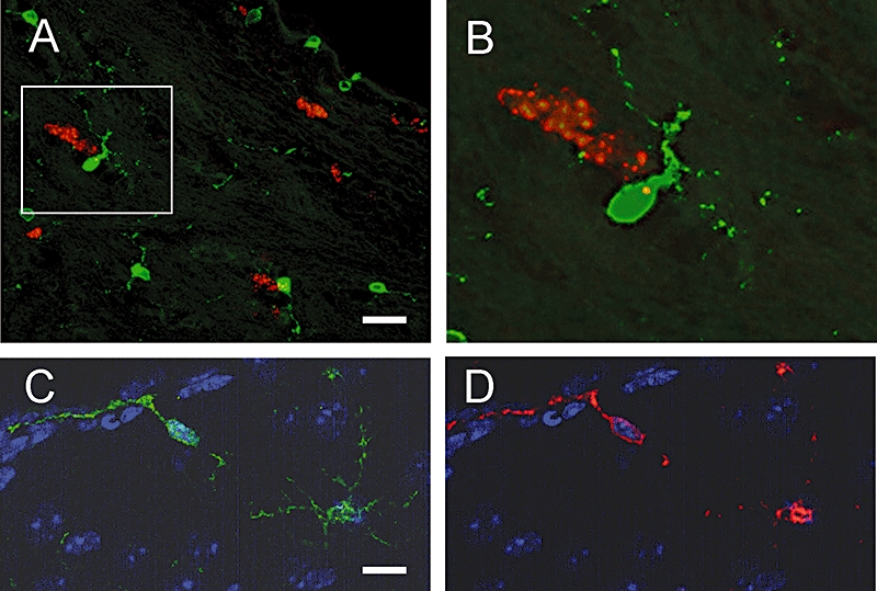
Figure 5.

Expression of COX-2 in the SVZ of the adult mouse brain. (A) and (B) Representative micrographs from coronal sections of the SVZ double stained for COX-2 (green) and Ki-67 (red). Box in (A) highlights a microglial cell. A higher magnification image of the cell is shown in (B). COX-2 was not seen in Ki-67-positive cells, but was found to be expressed in cells that closely associate with the proliferating cells. (C) and (D) Similar sections stained for COX-2 (green) and Iba1 (red), demonstrating that COX-2 is present in microglia around the ventricle. Scale bar in A is 40 µm, and 20 µm for (C) and (D).
