
Figure 2.
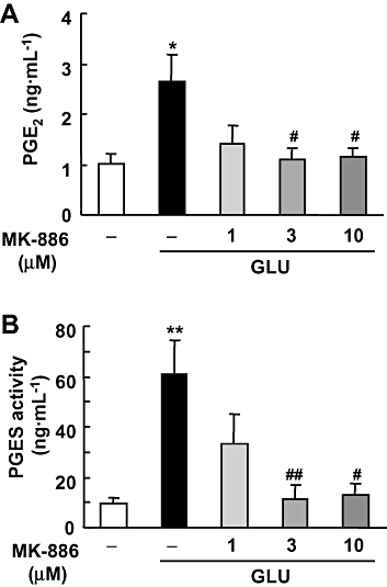
Effect of MK-886 on glutamate-induced PGE2 synthesis and PGES activity in cultures of rat hippocampal slices. MK-886 (1, 3 and 10 µM) was applied during and after the 1 mM glutamate (GLU) exposure for 15 min. (A) The amount of PGE2 in the culture medium 24 h after glutamate exposure was measured using an EIA kit (n = 4). (B) PGES activity in membrane fraction was measured as the conversion of exogenous PGH2 (0.5 µg) to PGE2 for 30 s by the lysate of these cells (30 µg protein). The amount of PGE2 was measured by the same protocol as in (A) (n = 4–5). **P < 0.01, *P < 0.05 versus the control; ##P < 0.01, #P < 0.05 versus glutamate alone. EIA, enzyme immunoassay; PGES, prostaglandin E synthase.