
Figure 6.
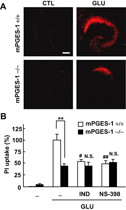
Effects of COX inhibitors on the glutamate-induced excitotoxicity in an mPGES-1 KO and a WT mouse hippocampal slice culture. PI uptake was analysed 24 h after 1 mM glutamate exposure for 30 min. (A) Representative confocal images of PI fluorescence of a control slice (CTL) and a slice that received glutamate (GLU) exposure in WT (+/+) mice and mPGES-1 KO (−/−) mice are shown (scale bar: 200 µm). (B) Summary data from PI uptake analysis in the CA1 region with or without glutamate and indomethacin (IND, 1 µM) or NS-398 (1 µM) were scaled to a percentage of the glutamate response in slices from WT mice. Indomethacin and NS-398 were applied during and after the glutamate exposure (n = 6–11 slices per group). **P < 0.01, ##P < 0.01, #P < 0.05, versus glutamate alone in a slice from a WT mouse; N.S. (not significant) versus glutamate alone in a slice from a mPGES-1 KO mouse. COX, cyclooxygenase; mPGES, microsomal prostaglandin E synthase; KO, knockout; PI, propidium iodide; WT, wild-type.