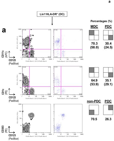
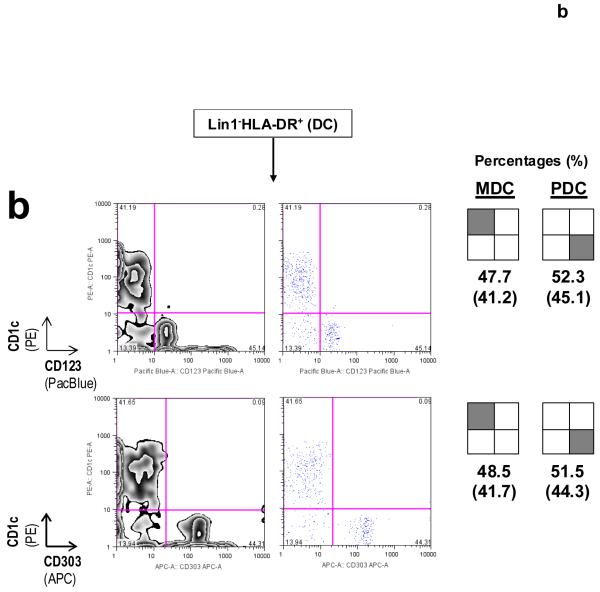
Figure 6.
DC subset-defining markers for identifying MDCs and PDCs as described in Protocol B. (a) CD11c and CD123, in conjunction with Lin1−HLA-DR+, are surface markers that are commonly used to identify MDCs and PDCs, respectively. CD1c+ (or BDCA-1+) MDCs might represent a major subset of CD11c+ MDCs, as reported previously30-32. Co-staining for CD123 and CD303 (BDCA-2) on Lin1−HLA-DR+ cells simultaneously identifies PDCs in the same staining reaction43; numbers in brackets represent raw percentages for non-PDCs (70.5%) and Lin1−HLA-DR+CD123+CD303+CD11c− PDCs (26.3%). Concordance between Lin1−HLA-DR+CD123+ and Lin1−HLA-DR+CD303+ cells is typically in the range 0.971 to 0.989 (mean 0.981, n = 6). (b) Immunostaining from another blood donor illustrates a variation in the MDC:PDC ratio or percentages. CD1c and CD303 can be used as alternative markers to identify specific Lin1−HLA-DR+ MDC and PDC subsets, respectively, and in determining subset ratios or percentages. Numbers inside brackets represent percentages calculated from all four quadrants, whereas percentages outside brackets are normalized for MDCs and PDCs only. Data are representative of at least three independent donors.


