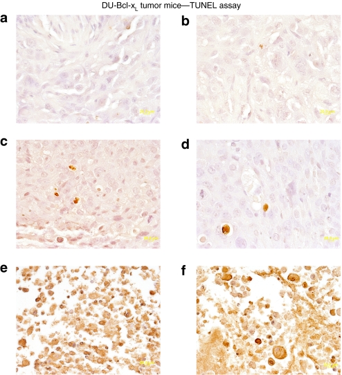
Figure 6.
Colorimetric TUNEL assay of therapy resistant DU-Bcl-xL tumor xenografts treated with microbubble/ultrasound (US)-guided Ad-GFP, Ad.mda-7, or cancer terminator virus (CTV). Subcutaneous tumor xenografts from DU-Bcl-xL cells were established in athymic nude mice in both right and left flanks, and only tumors on the right side were sonoporated following tail-vein injection of the indicated microbubble/Ad complexes during a course of 4 weeks. Tumors were removed fixed, sectioned, and stained to determine levels of double-stranded DNA breaks (TUNEL). Microscopy for TUNEL sections was under visible light at ×40 magnification (a representative of three separate tumors). (a,b) TUNEL staining of left and right side DU-Bcl-xL tumors following Ad-GFP/microbubble treatment. (c,d) TUNEL staining of left and right side DU-Bcl-xL tumors following Ad.mda-7-microbubble treatment. (e,f) TUNEL staining of left and right side DU-Bcl-xL tumors following CTV/microbubble treatment. Ad, adenovirus; GFP, green fluorescent protein; TUNEL, terminal deoxynucleotidyl transferase dUTP nick end labeling.

