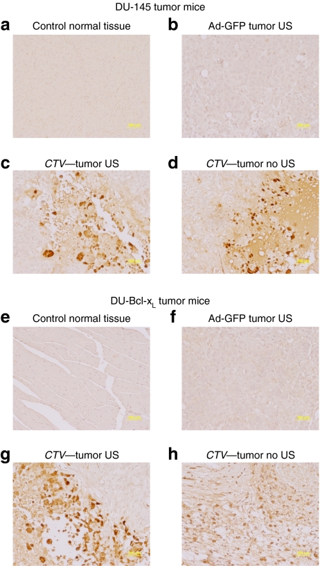
Figure 7.
Immunohistochemical analysis of DU-145 and therapy resistant DU-Bcl-xL tumor xenografts treated with microbubble/ultrasound (US)-guided Ad-GFP, Ad.mda-7, or cancer terminator virus (CTV). Subcutaneous tumor xenografts from DU-145 or DU-Bcl-xL cells were established in athymic nude mice in both right and left flanks, and only tumors on the right side were sonoporated following tail-vein injection of the indicated microbubble/Ad complexes during a course of 4 weeks. Tumors were removed fixed, sectioned, and immunostained to determine levels of E1A expression. (a) E1A immunohistochemical staining of control normal tissue from a DU-145 mouse treated with CTV/microbubble. (b) E1A immunohistochemical staining of control tumor tissues from a DU-145 mouse treated with Ad-GFP/microbubble and US. (c,d) E1A immunohistochemical staining of left and right side DU-145 tumors following CTV/microbubble treatment and US treatment of the tumor on the right side. (e) E1A immunohistochemical staining of control normal tissues from a DU-Bcl-xL mouse treated with CTV microbubbles. (f) E1A immunohistochemical staining of control tumor tissues from a DU-Bcl-xL mouse treated with Ad-GFP microbubbles and US. (g,h) E1A immunohistochemical staining of left and right side DU-Bcl-xL tumors following CTV/microbubble treatment of the tumor on the right side. Ad, adenovirus; GFP, green fluorescent protein.

