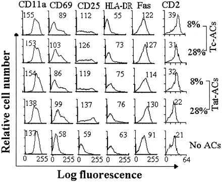
Figure 5.

Surface expression of activated antigens on PBT cells stimulated with immobilized anti-CD3 mAb in the presence of ACs. PBT cells were stimulated in the presence of indicated percentages of C-ACs, Tat-ACs, or Tc-ACs prepared as in Fig. 1. After incubation for 4 days at 37°C, the cells were stained by indicated mAbs. The mean fluorescent intensity for analysis of 10,000 events on T cell gate of each sample is indicated. Fluorescent profiles of PBT cells stimulated by immobilized anti-CD3 mAb in the absence of ACs are shown as controls.
