
Figure 6.
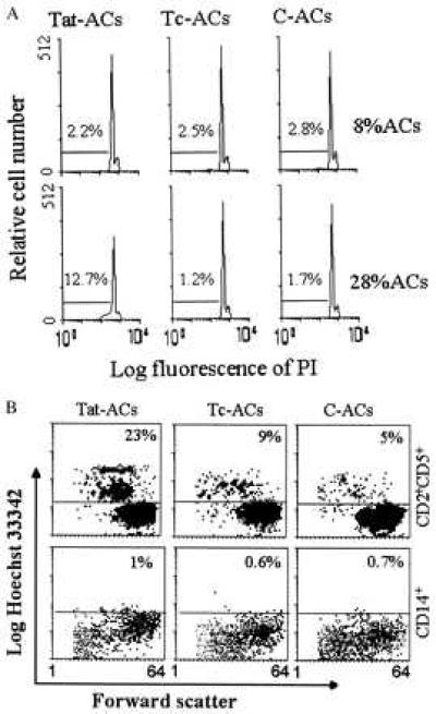
PI (A) and Hoechst (B) staining of apoptotic cell death in PBT cells stimulated with immobilized anti-CD3 mAb in the presence of ACs. (A) PBT cells were stimulated for 4 days at 37°C in the presence of either 8% or 28% of Tat-ACs, Tc-ACs or C-ACs, after which the cells were stained by PI and DNA contents were examined by flow cytometry. The percentages of cells identified as apoptotic cells (subG1 cell population) are indicated. (B) PBT cells were stimulated for 4 days at 37°C in the presence of 28% of indicated ACs, after which the cells were stained with FITC-conjugated anti-CD2 mAb plus phycoerythrin-conjugated anti-CD5 mAb, or with FITC-conjugated anti-CD14 mAb alone, followed by Hoechst staining. The percentages denote Hobright cells within CD2+CD5+ or CD14+ cell population.