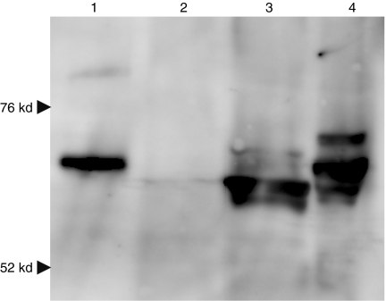
Figure 1.
Western blot analysis of Hsp70 and Hsp70-VP22268–301 expressed in B16-F10 cells. B16-F10 cells were transfected with pcDNA3.1 (2, mock), pHsp70 (3) or pHsp70-VP22268–301 (4) using Lipofectamine 2000, and cell lysates were subjected to 10% SDS-PAGE. After transfer to a membrane, Hsp70 and Hsp70-VP22268–301 were detected using mouse anti-Hsp70 monoclonal antibody. Lane 1 shows Hsp70-VP22268–301 expressed and purified from bacteria.

