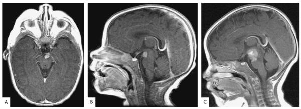
FIG. 2.
Magnetic resonance images of the infant with an atypical teratoid/rhabdoid tumor. A, B. Postcontrast T1 axial and sagittal MRIs at presentation shows a 6 × 8 × 8 mm mass with homogenous enhancement within the interpeduncular cistern inseparable from the left third cranial nerve. C. Postcontrast T1 sagittal MRI performed 1 month later shows an increase in the size of the mass to 22 × 18 × 18 mm with extension along the third cranial nerve and invasion of the midbrain and pons.

