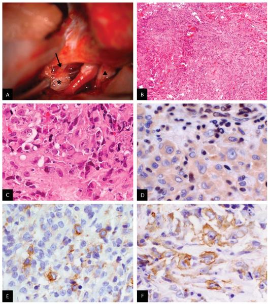
FIG. 3.
A. Intraoperative photograph: superior view of the anterior brainstem. The third cranial nerve (arrow) is lateral to the internal carotid artery, and the optic nerve (triangle) is located medially. A white, gelatinous tumor (star) is seen arising from the posterior aspect of the third cranial nerve. B–F. Histopathology of the surgical specimen. B. Small and large blue cells arranged in cords and nodules separated by fibrous septae with hemorrhage, necrosis, and mitotic figures (hematoxylin and eosin: original magnification ×40). C. Anaplastic, large pink cells with cytoplasmic inclusions and vesicular nuclei with prominent nucleoli (hematoxylin and eosin; original magnification: ×400). D. Large tumor cells exhibit no nuclear labeling for integrase interactor-1 (INI1), whereas infiltrating lymphocytes and endothelial cells show the expected nuclear reactivity of normal cell types (BAF47 anti-INI1 immunohistochemical staining; original magnification ×400) (4). E. Tumor cells exhibit positive labeling for epithelial membrane antigen (EMA) (anti-EMA immunohistochemical staining; original magnification: ×400). F. Tumor cells exhibit positive labeling for smooth muscle actin (SMA) (anti-SMA immunohistochemical staining; original magnification ×400).

