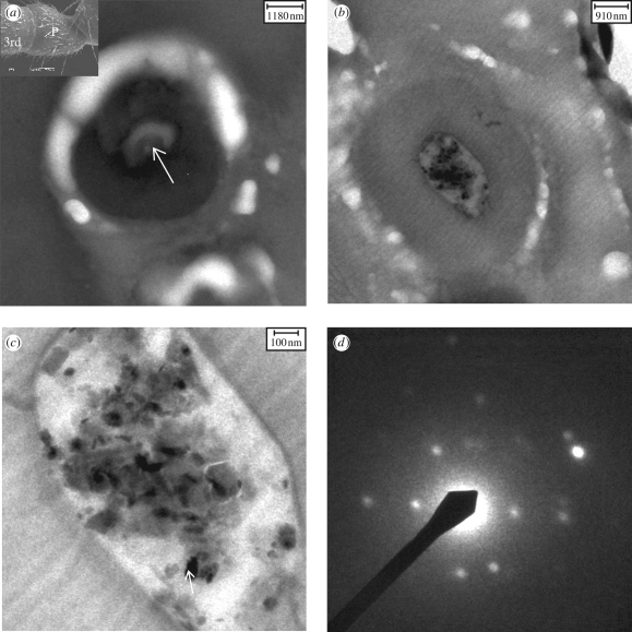
Figure 4.
Transmission electron microscopy micrographs of transversal ultrathin sections of the third segment/pedicel joint cut through the antenna. (a) A cuticular knob (diameter, 4–5 µm). The white arrow points to a long sensory process (diameter, 1 µm). Inset: SEM of P. marginata antenna, showing the third segment (3rd) and the pedicel (P). (b) Another cuticular knob in which particles were found. (c) Magnified knob region in which haematite and goethite (arrow) crystals together with silicates/alumosilicates were identified. (d) Diffraction pattern of the goethite particle (arrow in c).

