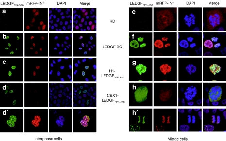
Figure 2.
Subcellular localization and HIV-IN interaction of stably expressed LEDGF hybrids in interphase and mitotic cells. Stable KD cell lines were complemented with LEDGF325–530 fusion proteins and transfected with mRFP-INs. Laser scanning microscopy (LSM) images of cells stained with anti-LEDGF325–530 antibody are shown (green). Nuclei were stained with DAPI (4',6-diamidino-2-phenylindole; blue). The respective constructs are indicated. Mitotic cells are displayed in the right panel. The data are representative for the vast majority of the imaged cells. CBX1, heterochromatin protein 1β (formerly HP1β); H1, histone H1; HIV, human immunodeficiency virus; IN, integrase; KD, knockdown; LEDGF, lens epithelium–derived growth factor; mRFP, monomeric red fluorescent protein; NLS, nuclear localization signal.

