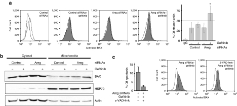
Figure 4.
Areg inactivates BAX in H358 cells. H358 cells were transfected with control or Areg siRNAs. As indicated, 0.5 µmol/l gefitinib and/or 10 µmol/l z-VAD-fmk were added. (a) Flow cytometry analysis of BAX immunostaining using activated-BAX antibody. Dotted histogram and IgG, irrelevant antibody; open histogram, control cells; filled histogram, treated cells as indicated. Percentages of activated-BAX-stained cells were expressed as mean ± SD (n ≥ 3). *P < 0.05, more significant than control. (b) H358 cells were fractionated into cytosolic and mitochondrial fractions. Both fractions extracts were subjected to western blotting using BAX antibody. Mitochondrial HSP70 was used for checking that cytosolic extracts were mitochondria-free and actin for loading control. (c) Apoptosis was scored after counting Hoechst-stained cells. Flow cytometry analysis of BAX immunostaining as described in a. Areg siRNAs, amphiregulin small-interfering RNAs.

