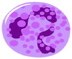
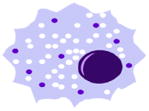
Table 1.
Effects of aging on phagocytic cell function*
| Functional status | Phagocytes | |
|---|---|---|
| Neutrophils | Macrophages | |
 |
 |
|
| Reduced | Receptor recruitment to lipid rafts Signal transduction (e.g., Ca2+ influx, phosphorylation of ERK, p38, Akt, PLC-γ) Chemotaxis Phagocytosis |
Signal transduction (e.g., total levels and/or activation of STAT-1α, p38 & JNK MAPKs, MyD88, NF-κB) Chemotaxis Cytokine production (IL-6, TNF-α, MIP-1α, MIP-1β, MIP-2) Reactive oxygen species production |
| Maintained | Total cell number Receptor expression (GM-CSFR, TLR2, TLR4, CD14, CD11b) Apoptosis (spontaneous) |
Expression of IFN-γR and TLRs (TLR2, TLR4, TLR5, TLR6) Phagocytic receptor expression (CD14, CD11b, CD18, CD36, mannose receptor [CD206], dectin-1, scavenger receptor-AI) Phagocytosis (?) Expression of TLR negative regulators (e.g., SOCS-1, IRAK-M, A20, PPAR-γ) |
| Increased | Apoptosis (under priming conditions) | Receptors involved in inflammation amplification (C5aR, TREM-1) PGE2 production |
| Controversial or both increase/decrease observed | Reactive oxygen species production | Nitric oxide production and intracellular killing TLR1 (decreased in human monocytes; unaltered in mouse macrophages) |
This compilation is based on a great number of studies, mainly using mouse or human cells, and original work has been cited in excellent specialized reviews (Butcher et al., 2000; Fulop et al., 2004; Gomez et al., 2008; Kovacs et al., 2009; Plowden et al., 2004; Solana et al., 2006; Stout and Suttles 2005).
