
Figure 7. Trib2 forms a complex with C/EBPα and results in its proteosomal-dependent degradation.
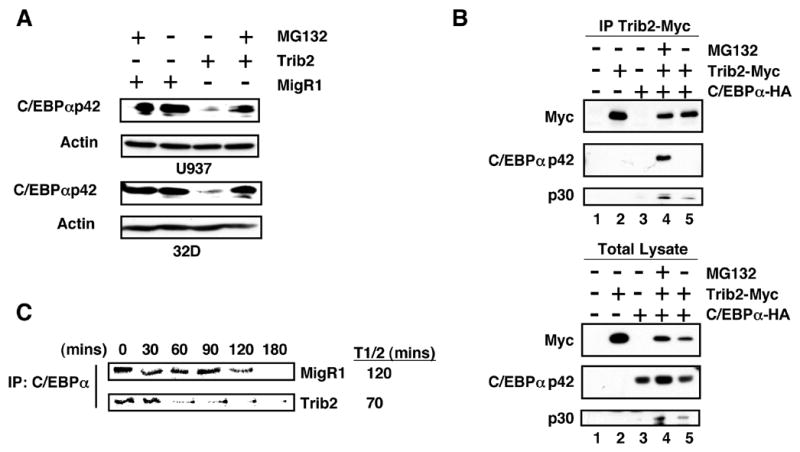
(A) Sorted GFP+ U937 (top panel) and 32D (lower panel) cells transduced with either MigR1 or Trib2, treated +/− 10 μM MG132 for 2 hrs were assessed for C/EBPα expression by western blot.
(B) 293T cells were transfected with empty vector (lane 1), myc-tagged Trib2 (lane 2), HA-tagged C/EBPα (lane 3), or co-transfected with both (lanes 4 and 5), and treated with 10 μM MG132 for 2 hrs (lane 4). Trib2 was immunoprecipitated using a Myc 9E10 antibody and western blotting performed with HA and Myc antibodies on immunoprecipitates (top panel) and total lysates (lower panel).
(C) U937 cells transduced with MigR1 or Trib2 were metabolically labeled with 35S-methionine for 60 min and chased for the indicated times. C/EBPα was immunoprecipitated and radiolabeled protein detected by SDS-PAGE. The half-life (T1/2) was calculated using ImageJ software.