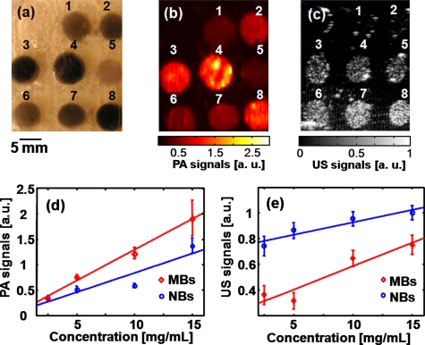
Figure 2.
(a) Photograph of a phantom containing tumor simulators made of encapsulated-ink MBs and NBs with various concentrations. (b) The corresponding PA image. (c) The corresponding US image. (d) The quantification of the PA signals at various concentrations of MBs and NBs. (e) The quantification of the US signals at various concentrations of MBs and NBs. 1 through 4: MBs at concentrations of 2.5, 5.0, 10, and 15 mg∕mL, respectively. 5 through 8: NBs at concentrations of 2.5, 5.0, 10, and 15 mg∕mL, respectively.

