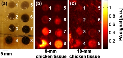
Figure 3.
(a) Photograph of a phantom containing tumor simulators made of encapsulated-ink MBs and NBs with various concentrations. (b) The corresponding PA image of the phantom positioned below 8 mm of chicken breast tissues. (c) The corresponding PA image of the phantom positioned below 18 mm of chicken breast tissues. 1 through 4: MBs at concentrations of 2.5, 5.0, 10, and 15 mg∕mL, respectively. 5 through 8: NBs at concentrations of 2.5, 5.0, 10, and 15 mg∕mL, respectively.

