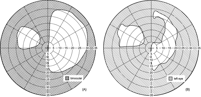
Figure 10.
Prism scotoma fitting. A peripheral prism for hemianopia creates visual field expansion (patient without the peripheral prism would only see to the right of the vertical midline). (a) Binocular visual field with an upper peripheral prism worn over the left eye. When probed binocularly, the fellow eye compensates (on the seeing side, here the right side) for the prism’s apical scotoma, so there is no reduction in the right portion of the visual field. (b) Monocular visual field of the left eye when stimuli are presented dichoptically so they were seen only by the prism eye: the effect of the apical scotoma is evident. A plot of this nature helps identify proper fitting of the peripheral prism. In this case, the peripheral prism could be moved to the right to reduce or eliminate the gap between the normal and expanded visual fields in binocular viewing. The fixation mark and background were presented binocularly in both plots. The difference in the extent of the visual field on the right side [ending at 30 deg in (a) and 20 deg in (b)] is due to the greater limitation from the frame of the goggles on the left eye than the right eye (available in the binocular test).

