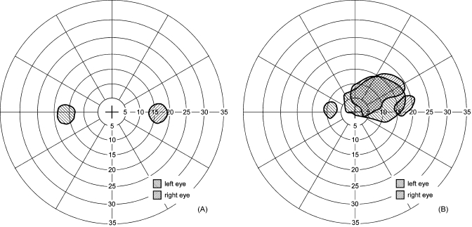
Figure 8.
Dichoptic visual fields. Monocular, right (right-tilted stripes) and left (left-tilted stripes) scotomas found in the visual fields when measured with our dichoptic visual test system while viewing binocularly for (a) a normally sighted subject, and (b) a patient with bilateral central scotomas. For the normally sighted subject, both physiological blind spots (optic nerve heads) are measured separately while the observer was fixating on a binocularly visible target and was seeing the physical screen binocularly. The patient used the same preferred retinal locus (PRL) for fixation here as when viewing monocularly with the right eye (i.e., not the left PRL). Note how the left eye central scotoma includes the fixation target. These visual fields illustrate the ability to measure each eye separately under binocular conditions.

