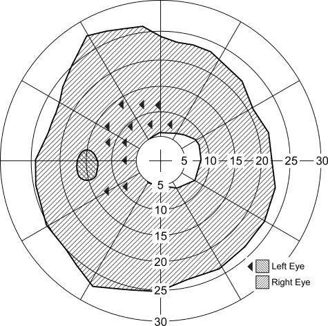
Figure 9.
Ring scotoma probe. Dichoptic visual field plot of a normally sighted subject fixating through a 3× bioptic telescope mounted on the right spectacle lens while the left eye was open. The clear central area represents the visual field visible to the right eye through the telescope, and the large hatched area is the ring scotoma caused by the 3× magnification of the telescope. The small cross-hatched area is not seen by either eye, as it is in the physiological blind spot of the left eye. The boundaries of those scotomas were found using kinetic stimuli. The left-pointing triangles represent locations at which static stimuli were presented only to the left eye. All of these static stimuli, presented within the ring scotoma of the right eye, were detected by the left eye. Under these conditions, vision in the left (nontelescope) eye was not suppressed, and therefore, objects appearing within the ring scotoma would be detected when viewing binocularly. The exception is the area of overlap of the ring scotoma with the blind spot, which is a (small) binocular scotoma.

