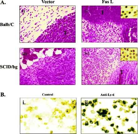
Figure 4.

Histological examination of the CT26-neo and CT26-FasL cells inoculated into BALB/c mice by hematoxylin/eosin staining. (A) (i) CT26-neo cells in BALB/c mice are shown 2 days after inoculation. The tumor cells (T) were growing intact. (ii) In CT26-FasL cells inoculated in BALB/c mouse, tumor cells were largely nonviable and an inflammatory cell infiltrate (Inset) was observed. (iii) CT26-neo cells in SCID-beige mouse. (iv) CT26-FasL cells in SCID-beige mouse. Infiltration of neutrophils (Inset) around the destroyed cancer cells was observed. (×400; Inset, ×1000.) (B) Immunostaining of inflammatory cells in a CT26-FasL tumor. The CT26-FasL tumor was dissected on day 2 and stained with an isotype control IgG (i) or anti-Ly-6G antibody (RB6–8C5) specific for neutrophils and activated monocytes/macrophages (ii) (18). A large percentage of the infiltrating cells in the CT26-FasL tumor were Ly-6G positive. (×400.)
