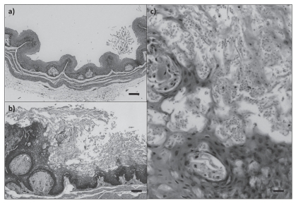
Figure 2.
Esophagi/crops of purple finches, Carpodacus purpureus. a) Normal tissue. Bar = 100 μm. b) Severe erosive and proliferative esophagitis/ingluvitis due to trichomoniasis. Note the intense staining of the basal layers that correspond to active replication (hyperplasia). Bar = 100 μm. c) Higher magnification of the lesion described in b, showing numerous protozoans (Trichomonas gallinae) among the keratinized epithelial layers. Bar = 20 μm.

