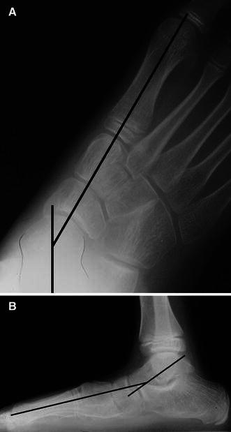
Fig. 7.

Standing radiographs of a flatfoot showing talus and first metatarsal axis lines crossing at the center of rotation of angulation (CORA) in the center of the head of the talus, indicating a single deformity at the talo-navicular joint. a Anteroposterior view. b Lateral view (Fig. 10-10, p. 144, from ref. [127], with permission)
