
Figure 10.
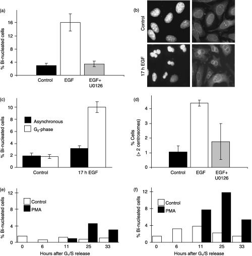
ERK activation during G2‐phase promotes hallmarks of chromosome instability; protective function of p21CIP1 expression. (a) Cells synchronized at the G1/S‐phase boundary were released for 7 h and treated with or without EGF (10 ng/mL) in the absence or presence of U0126 (10 µm). After 17 h, cells were immunostained for p62 nucleoporin as a marker of the nuclear envelope and counter‐stained with DAPI. The graph shows the percentage of bi‐nucleated cells under each condition. (b) Representative image of DAPI and p62 nucleoporin staining in control and EGF treated cells showing bi‐nucleated cells. (c) Percentage of bi‐nucleated cells in asynchronous or G2‐phase synchronized cells treated for 17 h in the absence or presence of EGF. (d) Percentage of cells containing greater than two centrosomes in control, EGF or EGF plus U0126 treated cells as described for (a). HCT116 parental (e) or p21 CIP1 knockout cells (f) were synchronized at G1/S phase, treated in the absence or presence of PMA at 6 h after release, and the percentage of bi‐nucleated cells were determined at the times indicated.