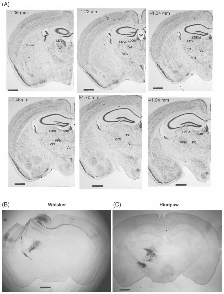
Figure 1.
Representative VL labeling following cortical injections. Representative Nissl stained coronal sections with thalamic motor nuclei identified, abbreviations as in Table I, numbers indicate relative position relative to bregma (Paxinos and Franklin 2001). Labeling following whisker (B) vs. hindpaw (C) identified different regions within VL. Scale bars represent 1 mm.

