
Fig. 3.
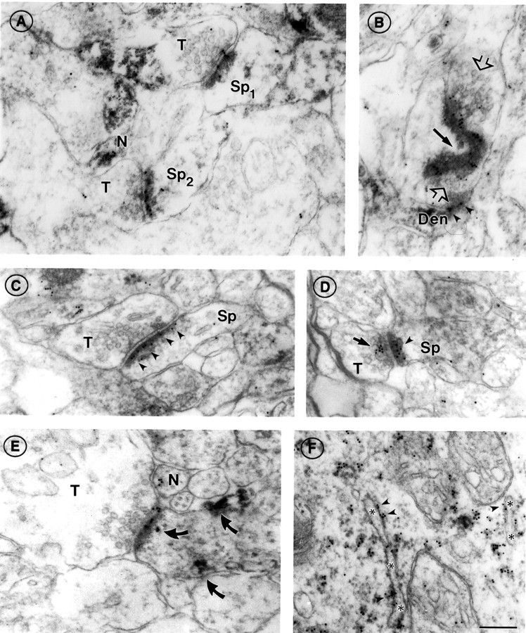
α7nAChR immunoreactivity at asymmetric synapses of PD 7 somatosensory cortex. A, Two axon terminals (T) forming synapses with spines (Sp1,Sp2) in layer I, showing α7nAChR labeling on the postsynaptic density (compare with Fig. 1). The top synapse also shows gold particles on the synaptic cleft and at the presynaptic membrane. Also note the cluster of α7nAChR labeling in the cytoplasm of an unidentified neurite (N).B, En face view of a large irregular α7nAChR-positive postsynaptic density in layers II/III. Black arrowpoints to the curvature of the PSD. The axon terminal, identifiable by synaptic vesicles (open arrows), converges on the PSD from two directions and appears to form another synapse below with a second dendritic compartment (Den), also α7nAChR positive (arrowheads). C,D, Axospinous synapses in layers II/III; compare the exclusively postsynaptic labeling in C(arrowheads) with the discrete clusters of presynaptic (arrow) and postsynaptic (arrowhead) labeling in D. The sparse, pleiomorphic vesicles seen in the axon terminal (T) in C are characteristic of immature synapses. E, An α7nAChR-positive synapse in layer VI. As in A, a nonsynaptic cluster of α7 label is seen in an adjacent neurite (N); also present in the postsynaptic compartment are patches of HRP–DAB reaction product indicating AMPAR labeling (curved arrows; compare with Fig. 1B; see also Fig. 6).A, D, and especially Eshow terminals with large cytoplasmic volumes devoid of vesicles. This is a recurrent feature of PD 7 neuropil. F, α7nAChR labeling of rough endoplasmic reticulum (ER) in a pyramidal cell body of layers II/III. Note the predominantly cytoplasmic location of gold particles (arrowheads; compare with Fig.1C). Asterisks denote the ER lumen. Scale bar (shown in F forA–F): 200 nm.