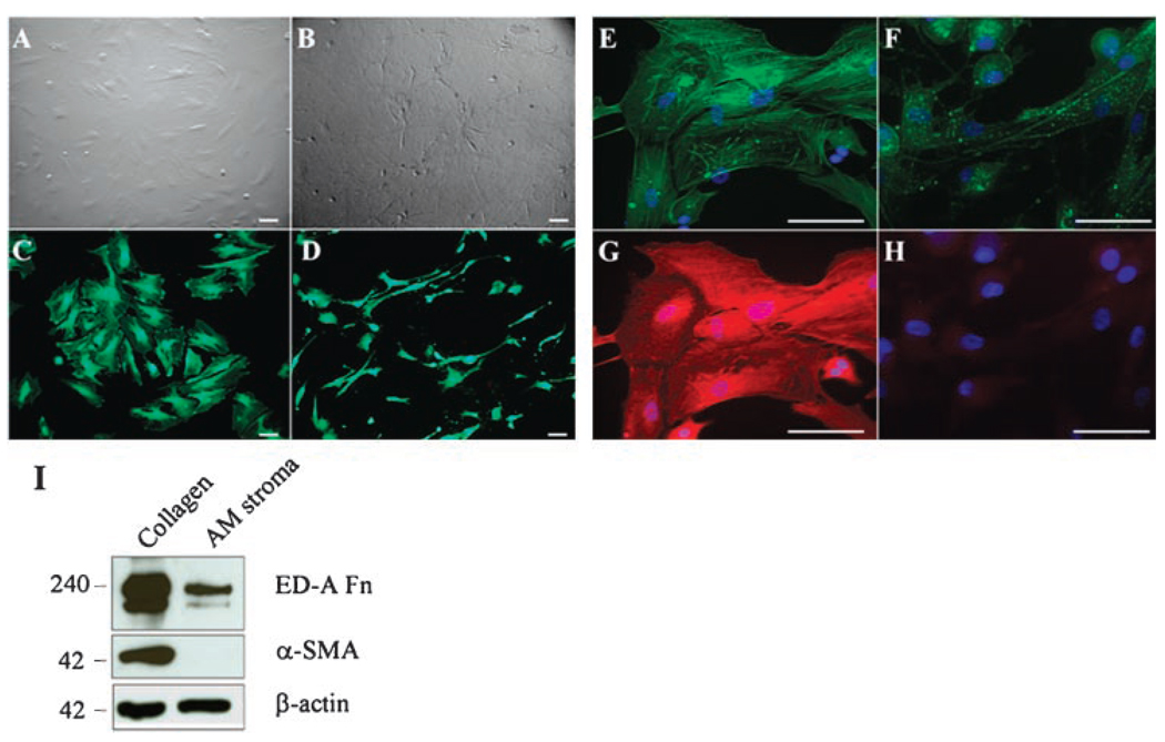
Fig 3.
Reversal of differentiated myofibroblasts to fibroblasts when AMSCs were cultured on AM stromal matrix. Myofibroblasts derived from AMSCs at P2 were subcultured on type I collagen (A,C,E,G) or AM stromal matrix (B,D,F,H) in DMEM with 10% FBS for 7 days. Live and Dead assay showed cells on both collagen (C) and AM stromal matrix (D) remained 100% viability, but exhibited a different cell shape. Phalloidin and α-SMA double staining showed vivid stress fibers (E) and strong α-SMA expression (G) in myofibroblasts on collagen cultures. In contrast, phalloidin staining became weak and spotty (F), and α-SMA became obscured in cells subcultured on AM stromal matrix (H). Western blot analysis showed decreased expression of ED-A fibronectin (Fn) and undetectable expression of α-SMA by AMSCs seeded on AM stromal matrix as compared to those seeded on type I collagen (I). Bars represent 100 µm.

