
Figure 2. Development of tissue inflammation driven by Th17 cells.
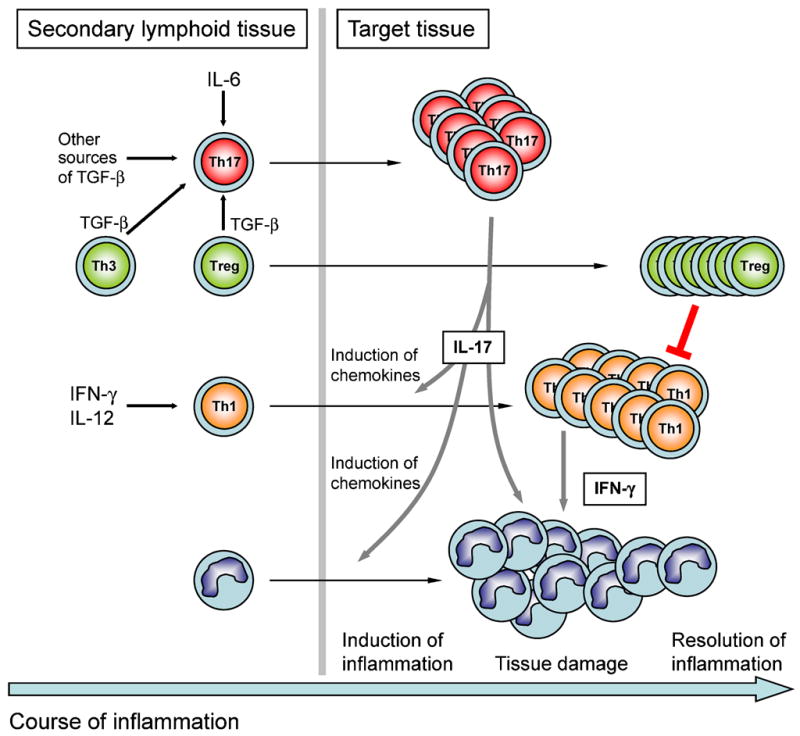
Antigen specific T cells are primed in secondary lymphoid tissue and driven into the Th17 developmental pathway in the presence of IL-6 and TGF-β. Sources of TGF-β are other T cells like Foxp3-expressing regulatory T cells (Treg) or Th3 cells. Similarly, antigen specific T cells are committed to the Th1 subset in the presence of IFN-γ and IL-12. Activated Th17 cells are among the first to infiltrate the target organ. By induction of chemokines, secondary waves of inflammatory cells like mononuclear cells and Th1 cells are attracted into the inflamed tissue. Both IL-17 produced by Th17 cells and IFN-γ produced by Th1 cells may act on myeloid cells to induce their effector functions that eventually lead to tissue damage. Whereas Th17 cells are prone to apoptosis, Th1 cells might finally be suppressed by T-reg cells that accumulate in the target organ and thus induce the resolution of inflammation.