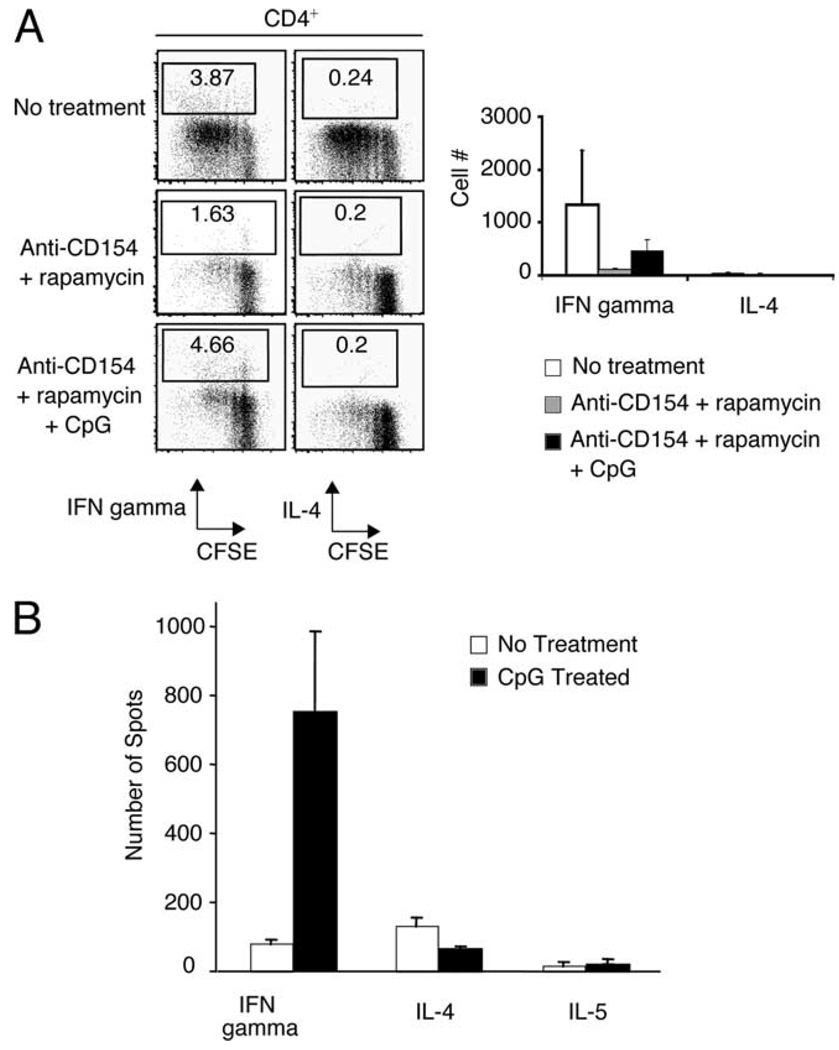
FIGURE 6.
CpG promotes Th1 differentiation from naive alloreactive T cells in vitro and in vivo. A, 1 × 10⁵ T-depleted bm12 splenocytes were cocultured for 96 h with 1 × 10⁵ CFSE-labeled CD4+CD25− T cells sorted from ABM mice. Anti-CD154 mAb (50 µg/ml), rapamycin (20 ng/ml), and CpG (3 µM) were added to individual culture wells. After 96 h of stimulation, the cells were washed and restimulated in vitro for intracellular cytokine analysis. Left panel, Frequency of CD4+IFN-γ and CD4+IL-4+ T cells in representative culture wells after 96 h. Right panel, Number of CD4+IFN-γ and CD4+IL-4+ T cells in culture wells after 96 h. Mean ± SD is shown of triplicate wells. Data are representative of two different experiments. B, B6 mice transplanted with bm12 cardiac allografts were treated with CpG (50 µg i.p.; days 0, 2, and 4) or PBS. Mice were subsequently sacrificed 2 wk after transplantation, and cytokine production from splenocytes from individual animals was assayed using an ELISPOT assay.

