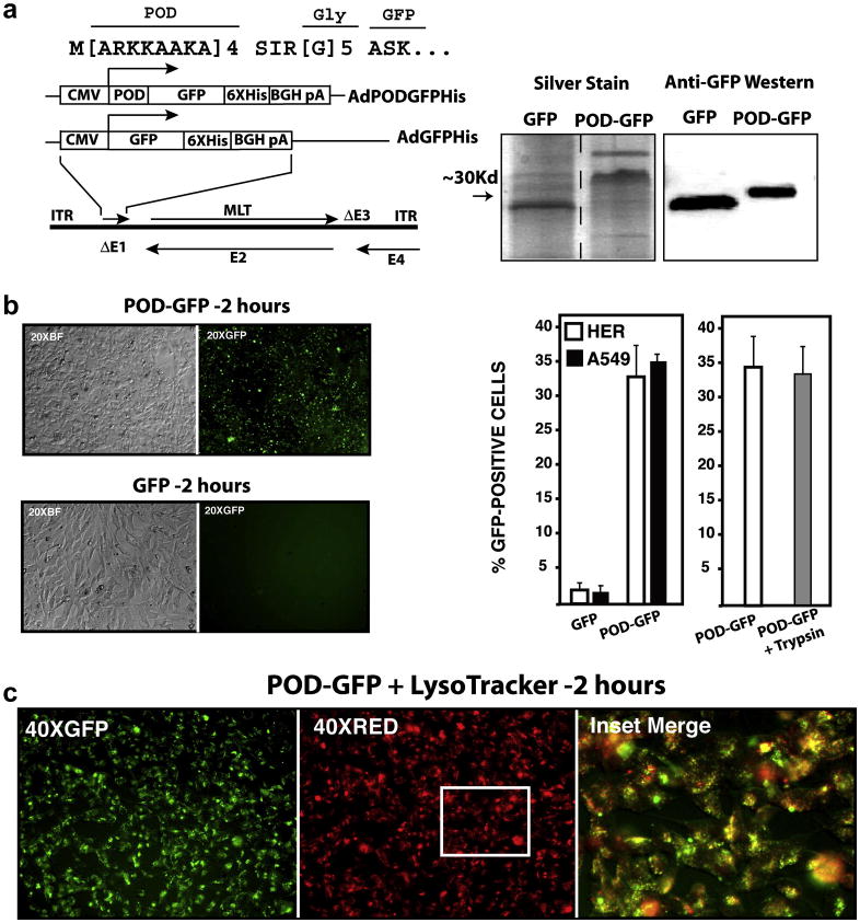
Figure 2. Recombinant adenovirus vectors expressing POD-GFP and uptake of POD-GFP into cells.
Expression cassettes coding for POD-GFP or GFP were cloned into the deleted E1 region (ΔE1) of a first generation adenovirus vector. POD was fused to GFP via a flexible polyglycine linker (a). Silver staining of purified protein indicated major bands of the anticipated molecular weights for POD-GFP and GFP. Anti-GFP western blot confirmed that these fusion proteins contained GFP (a). Purified POD-GFP or GFP protein was incubated with HER cells for 2 hours and GFP-positive cells were counted by FACS (b). POD-GFP entered cells and colocalized in part with Lysotracker, a marker of late endosomes (c). CMV, cytomegalovirus; 6XHis, His tag; BGH pA, bovine growth hormone polyadenylation signal; ITR, inverted terminal repeat; MLT, major late transcription unit; E1–E4; early regions 1–4 respectively.

