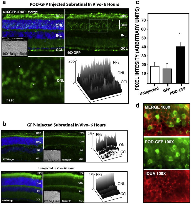
Figure 3. Transduction Properties of POD-GFP following subretinal injection in vivo.
POD-GFP or GFP protein was injected into the subretinal space of adult mice. POD-GFP entered the RPE and photoreceptor cell bodies in the ONL (a) and localized to photoreceptor cell body in a perinuclear, punctate and cytoplasmic manner (arrowheads). Surface plots of 40× retina indicate relative GFP-fluorescence units across the tissue. GFP-associated signal in GFP-injected eyes was not significantly above background auto fluorescence associated with uninjected eyes, except in the subretinal space as expected (b). Total fluorescence intensity associated with each experiment is quantified in (c). Lysosomal marker IDUA (red) indicated that POD-GFP (green) may not be sequestered in the lysosomes in vivo (d). ONL, Outer nuclear Layer; INL, Inner Nuclear Layer; GCL, Ganglion Cell Layer; RPE, Retinal Pigment Epithelium. Uninjected, n=3, GFP and POD-GFP, n = 4.

