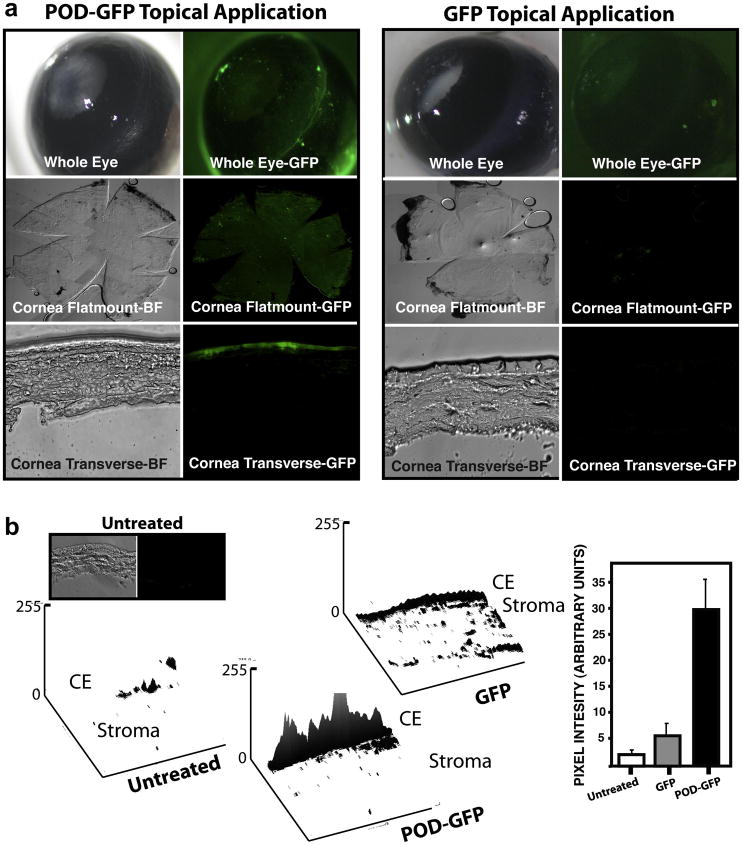
Figure 6. Topical application of POD-GFP on murine cornea in vivo.
Recombinant POD-GFP or GFP proteins were applied to the eyes of anesthetized adult mice and eyes washed and harvested 45 minutes later. Whereas GFP does not substantially bind to ocular tissues and is not internalized, POD-GFP binds to the entire corneal surface and can be readily detected in corneal wholemounts. Transverse sections of cornea indicates that POD-GFP localizes to the corneal epithelium. Surface plots of cross-sections indicates that the majority of the signal emanates from the corneal epithelium (b). CE, Corneal Epithelium; Untreated, n = 3, PODGFP and GFP, n = 4. Color version of this figure appears online.

