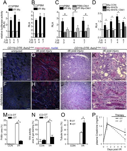
Fig. 2.
Macrophages are a source of Wnts and mediate Wnt responses in regeneration of the kidney epithelium. (A–D) Relative luciferase activity (RLA) from STF cells expressing Lrp5 or Lrp6 with Fzd3, Fzd4, or Fzd7, induced by coculture with: (A–C) d5 post-I/R kidney macrophages (I/R Mφ) compared with autologous peripheral blood monocytes (PBM) and inhibited (C) by recombinant Dkk1; or (D) coculture with bone marrow macrophages (Mφ-CON) or Wnt7b-expressing bone marrow macrophages and inhibited by the addition of Dkk1-expressing 293T cells. (E–L) PAS-stained kidney sections and F4/80 immunofluorescence confocal images of kidney sections from mice with and without conditional macrophage ablation during recovery from injury (asterisks, regenerating tubules; arrowheads, injured flattened epithelia; nec, necrotic debris). (M–O) Quantification of macrophages, active Wnt signaling (X-gal staining) in kidney cortex and medulla, and tubule injury d6 after injury. (P) Kidney function testing (plasma creatinine levels) in cohorts of mice (n = 6/group) with or without macrophage ablation from d3 to d6. Normal recovery is prevented by ablation. P < 0.05. n = 5 or 6/group. (Scale bars, 50 μm.)

