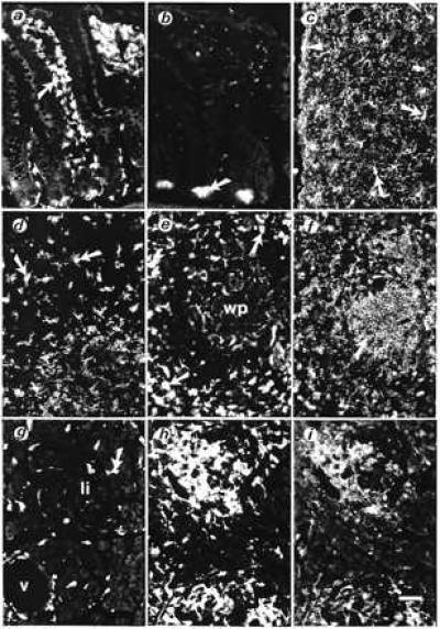
Figure 4.

Cellular localization of porcine daintain/AIF1-like immunoreactivity in tissues from normal BB-Wistar rats (a–e and g) and from diabetes-prone BB rats (without hyperglycemia, f, h, and i). (a) In sections of the ileum, strong daintain/AIF1-like immunoreactivity is found in a large number of dendritic cells beneath the epithelial layer (arrow), within the lamina propria of the tunica mucosa, and in scattered cells in the tunica submucosa. (b) Preabsorption of the daintain/AIF1 antibody with the purified peptide (10 μg/ml diluted antibody) greatly prevented the appearance of daintain/AIF1 immunoreactivity. Only autofluorescent cell profiles, such as Paneth cells (arrow), are present after absorption. (c) Daintain/AIF1-like immunoreactivity in ramified microglial cells (arrows) located over the entire cerebral cortex and concentrated beneath the pia mater (arrowhead). These cells also were labeled after incubation with an antibody OX-42 against CD11b/c antigen present in microglia. (d) Daintain/AIF1-like immunoreactivity in dendritic cells within the cortex of the thymus (arrows) and (e) in the spleen, present in scattered cells within the white pulp (wp) and abundantly in macrophages (arrows) of the red pulp. (g) Daintain/AIF1 immunoreactive cells scattered in the exocrine pancreas (arrow), mainly in the vicinity of vessels (v), and occasionally within pancreatic islets (Langerhan’s islets, li). (h) In prediabetic BB rats, strongly daintain/AIF1-like immunoreactive cells are found around and within pancreatic islets. (i) Daintain/AIF1 immunoreactivity colocalizes with MHC class II immunoreactivity in the same section of the pancreas of a prediabetic BB rat. In the spleen of the prediabetic BB rat, MHC class II immunoreactivity is present in lymphocytes of the white pulp (arrow, f), whereas daintain/AIF1 immunoreactivity, is more concentrated to the red pulp (compare with e). The magnification bar represents 50 μm, as shown in i.
