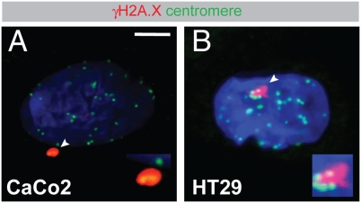
Fig. 6.
Centromere-associated DSBs in CIN cell lines. Caco2 (A) and HT29 (B) cells were seeded on glass coverslips, labeled with antibodies to γH2A.X (red) and centromeres (green), and then analyzed by confocal scanning microscopy. DNA was counterstained with DAPI (blue). Areas indicated with arrowheads are shown magnified twofold in Insets. Representative images are shown. (Scale bar, 5 μm.)

